Alternate header for print version


Licensing
Attribution Non-Commercial; No Derivatives:This image is licensed under a Creative Commons Attribution, Non-Commercial, No Derivatives License. View License Deed | View Legal Code
tweet  
*CIL – Cell Image Library accession number. Please use this to reference an image.

CIL:11434*  Cite 
Description

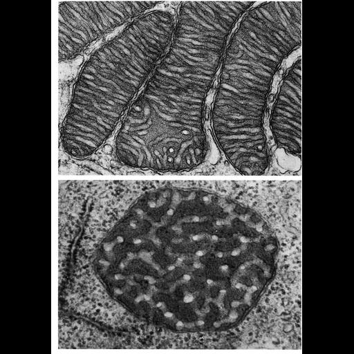
Figures 242 (upper) and 243 (lower) from Chapter 7 (Mitochondria) of 'The Cell, 2nd Ed.' by Don W. Fawcett M.D. Examples of tubular-shaped cristae from mitochondria in the zona fasciculata of hamster adrenal cortex (upper) and from the Singh amoeba (lower). Fig. 243 by Tom Pollard. A PDF copy of the accompanying chapter is available on the ASCB’s BioEDUCATE website.

Biological Sources
NCBI Organism Classification
Phodopus
Amoeba
Cellular Component
mitochondrion
mitochondrial crista
mitochondrial outer membrane
mitochondrial inner membrane
Biological Context
Biological Process
cellular respiration
Attribution
Names
Don W. Fawcett
Tom Pollard
Published
ISBN 0721635849
Link
Fawcett's "The Cell" 2nd Ed.
Citation
Digital Object Identifier (DOI)
doi:10.7295/W9CIL11434
Archival Resource Key (ARK)
ark:/b7295/w9cil11434
Grouping This image is part of a group.
Imaging
Image Type
transmission electron microscopy (TEM)
Image Mode
illumination by electrons
detection of electrons
Parameters Imaged
electron density
Source of Contrast
stain with broad specificity
Data Qualifiers
processed data
Sample Preparation
Methods
crosslinking-fixative fixed tissue
embedded tissue
Relation To Intact Cell
microtome-sectioned tissue
Dimensions
Spatial Axis Image Size Pixel Size
X 910px ——
Y 1284px ——