Alternate header for print version


Licensing
Attribution Non-Commercial; No Derivatives:This image is licensed under a Creative Commons Attribution, Non-Commercial, No Derivatives License. View License Deed | View Legal Code
tweet  
*CIL – Cell Image Library accession number. Please use this to reference an image.

CIL:11420*  Cite 
Description

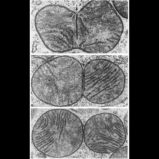
Figure 233 from Chapter 7 (Mitochondria) of 'The Cell, 2nd Ed.' by Don W. Fawcett M.D. Capturing stages of mitochondrial division in cells from the gastric mucosa of a mole with the electron microscope. A) The free edge of the developing septum has not yet reached the opposite side of the organelle. B) Asymmetrical penetration of the fold of the outer membrane is evident. C)Separation of the daughter mitochondrion is complete. Images by Toku Kanaseki. A PDF copy of the accompanying chapter is available on the ASCB’s BioEDUCATE website.

Biological Sources
NCBI Organism Classification
Talpidae
Cellular Component
mitochondrion
mitochondrial crista
mitochondrial outer membrane
mitochondrial inner membrane
Biological Context
Biological Process
mitochondrial fission
Attribution
Names
Toku Kanaseki
Published
ISBN 0721635849
Link
Fawcett's "The Cell" 2nd Ed.
Citation
Digital Object Identifier (DOI)
doi:10.7295/W9CIL11420
Archival Resource Key (ARK)
ark:/b7295/w9cil11420
Grouping This image is part of a group.
Imaging
Image Type
transmission electron microscopy (TEM)
Image Mode
illumination by electrons
detection of electrons
Parameters Imaged
electron density
Source of Contrast
stain with broad specificity
Data Qualifiers
processed data
Sample Preparation
Methods
crosslinking-fixative fixed tissue
embedded tissue
Relation To Intact Cell
microtome-sectioned tissue
Dimensions
Spatial Axis Image Size Pixel Size
X 807px ——
Y 1292px ——