Alternate header for print version


Licensing
Attribution Non-Commercial; No Derivatives:This image is licensed under a Creative Commons Attribution, Non-Commercial, No Derivatives License. View License Deed | View Legal Code
tweet  
*CIL – Cell Image Library accession number. Please use this to reference an image.

CIL:11417*  Cite 
Description

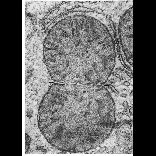
Figure 231 from Chapter 7 (Mitochondria) of 'The Cell, 2nd Ed.' by Don W. Fawcett M.D. Dividing mitochondrion in the rat liver. A septum of two membranes forms from the inner membrane. The outer mitochondrial membrane then invades the space between the septal membranes. When the advancing edges meet and fuse, the separation of the daughter mitochondria is completed. Image by Daniel Friend. A PDF copy of the accompanying chapter is available on the ASCB’s BioEDUCATE website.

Biological Sources
NCBI Organism Classification
Rattus
Cell Type
hepatocyte
Cellular Component
mitochondrion
mitochondrial crista
mitochondrial outer membrane
mitochondrial inner membrane
Biological Context
Biological Process
mitochondrial fission
Attribution
Names
Daniel Friend
Published
ISBN 0721635849
Link
Fawcett's "The Cell" 2nd Ed.
Citation
Digital Object Identifier (DOI)
doi:10.7295/W9CIL11417
Archival Resource Key (ARK)
ark:/b7295/w9cil11417
Grouping This image is part of a group.
Imaging
Image Type
transmission electron microscopy (TEM)
Image Mode
illumination by electrons
detection of electrons
Parameters Imaged
electron density
Source of Contrast
stain with broad specificity
Data Qualifiers
processed data
Sample Preparation
Methods
crosslinking-fixative fixed tissue
embedded tissue
Relation To Intact Cell
microtome-sectioned tissue
Dimensions
Spatial Axis Image Size Pixel Size
X 918px ——
Y 1288px ——