Alternate header for print version


Licensing
Attribution Non-Commercial; No Derivatives:This image is licensed under a Creative Commons Attribution, Non-Commercial, No Derivatives License. View License Deed | View Legal Code
tweet  
*CIL – Cell Image Library accession number. Please use this to reference an image.

CIL:11385*  Cite 
Description

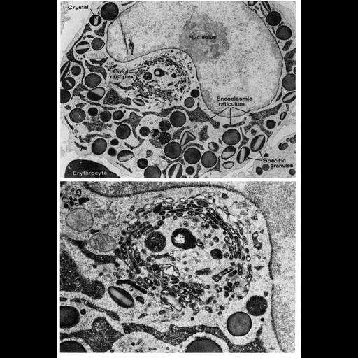
Figure 215 (upper) and 216 (higher magnification, lower) from Chapter 6 (Golgi Apparatus) of 'The Cell, 2nd Ed.' by Don W. Fawcett M.D. Granules of the eosinophilic myelocyte from rat bone marrow stained by the cytochemical reaction for peroxidase. In the lower panel, reaction product is evident in the cisternae of the Golgi, in condensing vacuoles and immature granules. Image from Bainton and Farquhar (1970) J. Cell Biol. 45:54-73, reprinted with permission. A PDF copy of the accompanying chapter is available on the ASCB’s BioEDUCATE website.

Biological Sources
NCBI Organism Classification
Rattus
Cell Type
eosinophilic myelocyte
Cellular Component
Golgi apparatus
Golgi stack
nucleus
nucleolus
endoplasmic reticulum
Attribution
Names
D. Bainton
Marilyn G Farquhar
Published
ISBN 0721635849
Pubmed
PMID:5459000
Link
http://www.ascb.org/bioeducate/FawcettTheCell...
http://jcb.rupress.org/content/45/1/54.long
Citation
Digital Object Identifier (DOI)
doi:10.7295/W9CIL11385
Archival Resource Key (ARK)
ark:/b7295/w9cil11385
Grouping This image is part of a group.
Imaging
Image Type
transmission electron microscopy (TEM)
Image Mode
illumination by electrons
detection of electrons
Parameters Imaged
electron density
Source of Contrast
stain with broad specificity
Data Qualifiers
processed data
Sample Preparation
Methods
crosslinking-fixative fixed tissue
embedded tissue
Relation To Intact Cell
microtome-sectioned tissue
Dimensions
Spatial Axis Image Size Pixel Size
X 871px ——
Y 1280px ——