Alternate header for print version


Licensing
Attribution Non-Commercial; No Derivatives:This image is licensed under a Creative Commons Attribution, Non-Commercial, No Derivatives License. View License Deed | View Legal Code
tweet  
*CIL – Cell Image Library accession number. Please use this to reference an image.

CIL:11362*  Cite 
Description

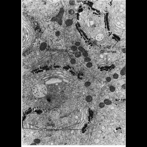
Figure 199 from Chapter 6 (Golgi Apparatus) of 'The Cell, 2nd Ed.' by Don W. Fawcett M.D. The Golgi apparatus in epithelial cells from the mouse epididymis. The convex, or cis, side of the Golgi shows selective deposition of osmium, whereas the concave, or trans, side is resistant to osmium staining even after long periods of postosmication. Tissue shown here was prepared with collidine-buffered osmium fixation followed by 40 hours postosmication at 37° C. Image by Daniel Friend. A PDF copy of the accompanying chapter is available on the ASCB’s BioEDUCATE website.

Biological Sources
NCBI Organism Classification
Mus musculus
Cell Type
epithelial cell
Cellular Component
Golgi apparatus
Golgi stack
mitochondrion
Golgi cis cisterna
Golgi trans cisterna
Attribution
Names
Daniel Friend
Published
ISBN 0721635849
Link
http://www.ascb.org/bioeducate/FawcettTheCell...
Citation
Digital Object Identifier (DOI)
doi:10.7295/W9CIL11362
Archival Resource Key (ARK)
ark:/b7295/w9cil11362
Grouping This image is part of a group.
Imaging
Image Type
transmission electron microscopy (TEM)
Image Mode
illumination by electrons
detection of electrons
Parameters Imaged
electron density
Source of Contrast
stain with broad specificity
Visualization Methods
osmium tetroxide
Data Qualifiers
processed data
Dimensions
Spatial Axis Image Size Pixel Size
X 902px ——
Y 1268px ——