Licensing

Attribution_by_nc_nd
Attribution Non-Commercial; No Derivatives:This image is licensed under a Creative Commons Attribution, Non-Commercial, No Derivatives License. View License Deed | View Legal Code
Comments: Powered by Pnyxe

Description

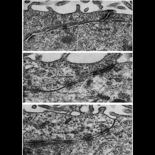
These examples of ependymal epithelium from the rat brain represent epithelia that lack zonulae occludentes. Further, there appears to be no consistent order to the junctional specializations that are present between these cells. Images courtesy of Enrico Mugnaini, are Figure 73 (upper), 74 (middle), 75 (lower) from Chapter 3 (Junctional Specializations) of 'The Cell, 2nd Ed.' by Don W. Fawcett M.D. A PDF copy of the accompanying chapter is available on the ASCB's BioEDUCATE website. A PDF copy of the accompanying chapter is available on the ASCB's BioEDUCATE website.

Biological Sources

NCBI Organism Classification
Rattus
(Old World rats)
Cell Type
ependymal cell
epithelial cell
Cellular Component
zonula adherens
gap junction
desmosome

Attribution

Name
Enrico Mugnaini
Published
ISBN 0721635849
Link
The Cell, 2nd Ed. by D.W. Fawcett

Grouping

This image is part of a group.

Imaging

Image Type
transmission electron microscopy (TEM)
Imaging Mode
illumination by electrons
detection of electrons
Parameters Imaged
electron density
Source of Contrast
stain with broad specificity
Visualization Methods
osmium tetroxide
Data Qualifiers
processed data

Dimensions

Spatial Axis Image Size Pixel Size
X 900px ——
Y 1260px ——
*CIL – Cell Image Library accession number. Please use this to reference an image.