Licensing

Attribution_by_nc_nd
Attribution Non-Commercial; No Derivatives:This image is licensed under a Creative Commons Attribution, Non-Commercial, No Derivatives License. View License Deed | View Legal Code
Comments: Powered by Pnyxe

Description

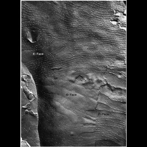
A freeze fracture replica of a Sertoli cell junction from rat testis shows more than 50 rows of particles along the E-face. Image from Gilula, Fawcett and Aoki, Dev. Biol. 50: 142-168 (1976), reprinted with permission as Figure 72 from Chapter 3 (Junctional Specializations) of 'The Cell, 2nd Ed.' by Don W. Fawcett M.D. A PDF copy of the accompanying chapter is available on the ASCB's BioEDUCATE website.

Biological Sources

NCBI Organism Classification
Rattus
(Old World rats)
Cell Type
Sertoli cell
Cellular Component
occluding junction
tight junction

Biological Context

Biological Process
cell-cell adhesion
cell-cell junction organization

Attribution

Names
Don W. Fawcett
Norton Gilula
Published
ISBN 0721635849
Links
The Cell, 2nd Ed. by D.W. Fawcett
Gilula et al. (1976) Dev. Biol. 50:142...
Pubmed
1269826

Grouping

This image is part of a group.
Data Qualifiers
processed data

Sample Preparation

Methods
freeze_fracture/freeze_etch
Relation To Intact Cell
isolated subcellular component

Dimensions

Spatial Axis Image Size Pixel Size
X 894px ——
Y 1236px ——
*CIL – Cell Image Library accession number. Please use this to reference an image.