Licensing
Attribution Non-Commercial; No Derivatives:This image is licensed under a Creative Commons Attribution, Non-Commercial, No Derivatives License. View License Deed | View Legal Code
Comments:
Powered by Pnyxe
Description
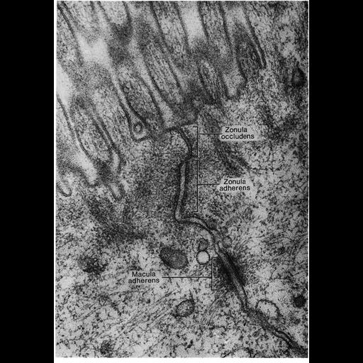
Electron micrograph of the junctional complex of intestinal epithelial cells of the rat shows the apical-most zonula occludens (tight junction), the zonula adherens (medium junction) and the macula adherens (desmosome). Image published in Faquhar and Palade, J. Cell Biol. 17: 375-412, 1963 and reprinted with permission as Figure 61 from Chapter 3 (Junctional Specializations) of 'The Cell, 2nd Ed.' by Don W. Fawcett M.D. A PDF copy of the accompanying chapter is available on the ASCB's BioEDUCATE website. A PDF copy of the accompanying chapter is available on the ASCB's BioEDUCATE website.
Biological Sources
- NCBI Organism Classification
- Rattus
- (Old World rats)
- Cell Type
- epithelial cell
- Cellular Component
- apical junction complex
- zonula adherens
- apical part of cell
- desmosome
Biological Context
- Biological Process
- maintenance of apical/basal cell polarity
Attribution
- Names
- Marilyn G. Farquhar
- George E. Palade
- Published
- ISBN 0721635849
- Links
- The Cell, 2nd Ed. by D.W. Fawcett
- Farquhar & Palade (1963). J. Cell Biol...
- Pubmed
- 13944428
Grouping
This image is part of a group.Imaging
- Image Type
- transmission electron microscopy (TEM)
- Imaging Mode
- illumination by electrons
- detection of electrons
- Parameters Imaged
- electron density
- Source of Contrast
- stain with broad specificity
- Visualization Methods
- osmium tetroxide
- Data Qualifiers
- processed data
Sample Preparation
- Methods
- crosslinking-fixative fixed tissue
- tissue in plastic embedment
- Relation To Intact Cell
- microtome-sectioned tissue
Dimensions
| Spatial Axis | Image Size | Pixel Size |
|---|---|---|
| X | 898px | —— |
| Y | 1276px | —— |
*CIL – Cell Image Library accession number. Please use this to reference an image.