Licensing

Attribution_by_nc_nd
Attribution Non-Commercial; No Derivatives:This image is licensed under a Creative Commons Attribution, Non-Commercial, No Derivatives License. View License Deed | View Legal Code
Comments: Powered by Pnyxe

Description

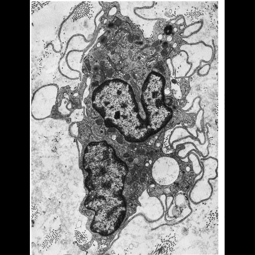
A thin section through a macrophage from interstitial tissue of the testes of the opossum. These cells are characterized by extensive folding of the cell surface, some of which appear to have captured fluid from the extracellular environment through pinocytosis. Figure 50 from Chapter 2 (Specializations of the Free Surface) of 'The Cell, 2nd Ed.' by Don W. Fawcett M.D. A PDF copy of the accompanying chapter is available on the ASCB's BioEDUCATE website.

Biological Sources

NCBI Organism Classification
Didelphimorphia
(American marsupials)
Cell Type
macrophage
Cellular Component
lysosome
nucleus

Biological Context

Biological Process
pinocytosis
endocytosis

Attribution

Name
Don W. Fawcett
Published
ISBN 0721635849
Link
Fawcett's "The Cell, 2nd Ed."

Grouping

This image is part of a group.

Imaging

Image Type
transmission electron microscopy (TEM)
Imaging Mode
illumination by electrons
detection of electrons
Parameters Imaged
electron density
Source of Contrast
stain with broad specificity
Visualization Methods
osmium tetroxide
Data Qualifiers
processed data

Sample Preparation

Methods
crosslinking-fixative fixed tissue
Relation To Intact Cell
microtome-sectioned tissue

Dimensions

Spatial Axis Image Size Pixel Size
X 918px ——
Y 1200px ——
*CIL – Cell Image Library accession number. Please use this to reference an image.