Licensing

Attribution_by_nc_nd
Attribution Non-Commercial; No Derivatives:This image is licensed under a Creative Commons Attribution, Non-Commercial, No Derivatives License. View License Deed | View Legal Code
Comments: Powered by Pnyxe

Description

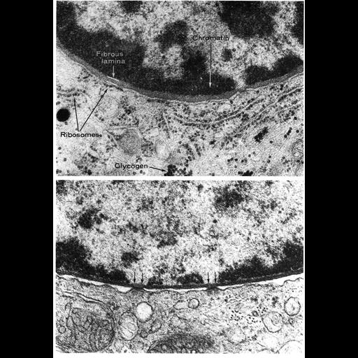
Upper panel shows a transmission electron micrograph featuring the nuclear envelope with the nuclear lamina seen here as a uniform layer between the inner nuclear membrane and more darkly staining blocks of heterochromatin in this human skin cell. The lamina appears to be discontinuous over the nuclear pore. Lower panel shows a thinner and more darkly staining lamina in a rodent Leydig cell. Figures 157 (upper, courtesy of George Szabo) and 158 from Chapter 4 (Nucleus) of 'The Cell, 2nd Ed.' by Don W. Fawcett M.D. A PDF copy of the accompanying chapter is available on the ASCB's BioEDUCATE website.

Biological Context

Biological Process
nucleus organization
nuclear transport
chromatin organization

Attribution

Names
George Szabo
Don W. Fawcett
Publisheds
ISBN 0721635849
from: Don W. Fawcett M.D ‘The Cell' 2nd edition, W.B. Saunders Chapter 4 (Nucleus)
Link
ASCB BioEDUCATE Program

Grouping

This image is part of a group.

Imaging

Image Type
film
Imaging Mode
transmission electron microscopy (TEM)
detection of electrons
Parameters Imaged
electron density
Source of Contrast
differences in adsorption or binding of stain
Visualization Methods
uranyl salt
osmium tetroxide
lead salt
Processing History
Image extracted from .pdf file of 'The Cell'
Data Qualifiers
processed data

Dimensions

Spatial Axis Image Size Pixel Size
X 898px ——
Y 1276px ——
*CIL – Cell Image Library accession number. Please use this to reference an image.