Alternate header for print version


Licensing
Attribution Non-Commercial; No Derivatives:This image is licensed under a Creative Commons Attribution, Non-Commercial, No Derivatives License. View License Deed | View Legal Code
tweet  
*CIL – Cell Image Library accession number. Please use this to reference an image.

CIL:11029*  Cite 
Description

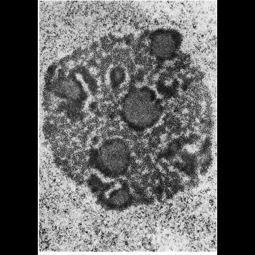
Transmission electron micrograph of a cross section through the nucleolus from a Chinese hamster spermatogonium revealing a complex distribution of regions of varying granular textures.

Technical Details

This micrograph, courtesy of David Phillips, was published as Figure 134 from Chapter 4 (Nucleus) of 'The Cell, 2nd Ed.' by Don W. Fawcett M.D. A PDF copy of the corresponding chapter is available on the ASCB's BioEDUCATE website.

Biological Sources
NCBI Organism Classification
Cricetulus griseus
Cell Type
spermatogonium
Cellular Component
nucleolus
nucleus
nuclear chromatin
Biological Context
Biological Process
nucleus organization
RNA metabolic process
nucleolus organization
Attribution
Names
David Phillips
Published
ISBN 0721635849
Published
from: Don W. Fawcett M.D ‘The Cell’ 2nd edition, W.B. Saunders Chapter 4 (Nucleus)
Link
ASCB BioEDUCATE Program
OTHER
Don W Fawcett
Citation
Digital Object Identifier (DOI)
doi:10.7295/W9CIL11029
Archival Resource Key (ARK)
ark:/b7295/w9cil11029
Grouping This image is part of a group.
Imaging
Image Type
film
Image Mode
transmission electron microscopy (TEM)
detection of electrons
Parameters Imaged
electron density
Source of Contrast
differences in adsorption or binding of stain
Visualization Methods
uranyl salt
osmium tetroxide
lead salt
Processing History
Image extracted from .pdf file of 'The Cell'
Data Qualifiers
processed data
Dimensions
Spatial Axis Image Size Pixel Size
X 908px ——
Y 1292px ——