Licensing

Attribution_by_nc_nd
Attribution Non-Commercial; No Derivatives:This image is licensed under a Creative Commons Attribution, Non-Commercial, No Derivatives License. View License Deed | View Legal Code
Comments: Powered by Pnyxe

Description

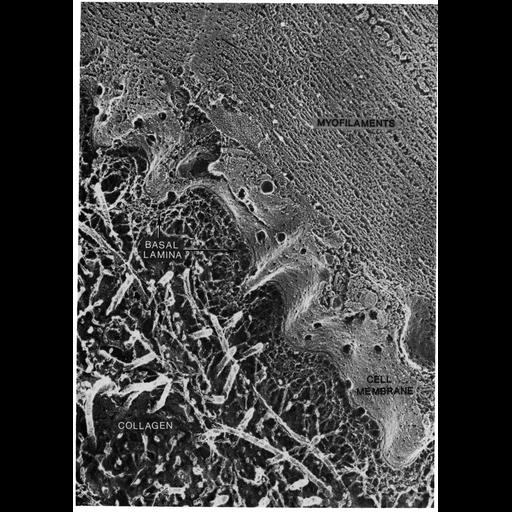
Quick-frozen deep-etched preparation of skeletal muscle tissue shows the muscle fiber and associated extracellular components. The basement membrane (labeled basal lamina) lies adjacent to the undulating sarcolemma. Myofilaments of the muscle fiber are composed of actin and myosin filaments. Image by J. Heuser, Figure 30 from Chapter 1 (The Cell Surface) of 'The Cell, 2nd Ed.' by Don W. Fawcett M.D. A PDF copy of the accompanying chapter is available on the ASCB's BioEDUCATE website.

Attribution

Name
John Heuser
Published
ISBN 0721635849
Link
The Cell, 2nd Ed. by D.W. Fawcett

Grouping

This image is part of a group.
Data Qualifiers
processed data

Sample Preparation

Methods
freeze_fracture/freeze_etch
Relation To Intact Cell
isolated subcellular component
*CIL – Cell Image Library accession number. Please use this to reference an image.