Alternate header for print version


Licensing
Attribution Non-Commercial; No Derivatives:This image is licensed under a Creative Commons Attribution, Non-Commercial, No Derivatives License. View License Deed | View Legal Code
tweet  
*CIL – Cell Image Library accession number. Please use this to reference an image.

CIL:10948*  Cite 
Description

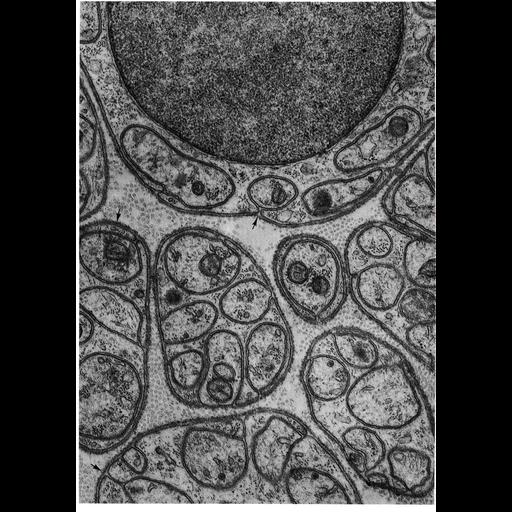
Electron micrograph of a nerve from the mesentery of a rat shows groups of unmyelinated axons wrapped by deeply invaginating Schwann cells in cross section. Surrounding the Schwann cell wrapping is a secreted basement membrane (known as the lamina externa, or boundary layer), similar to the basal lamina produced by epithelial cells. Figure 25 from Chapter 1 (The Cell Surface) of 'The Cell, 2nd Ed.' by Don W. Fawcett M.D. A PDF copy of the accompanying chapter is available on the ASCB's BioEDUCATE website.

Biological Sources
NCBI Organism Classification
Rattus
Cell Type
myelinating Schwann cell
peripheral neuron
Cellular Component
basement membrane
extracellular region part
plasma membrane
axon
Attribution
Names
Don W. Fawcett
Published
ISBN 0721635849
Link
The Cell, 2nd Ed. by D.W. Fawcett
Citation
Digital Object Identifier (DOI)
doi:10.7295/W9CIL10948
Archival Resource Key (ARK)
ark:/b7295/w9cil10948
Grouping This image is part of a group.
Imaging
Image Type
transmission electron microscopy (TEM)
Image Mode
illumination by electrons
detection of electrons
Parameters Imaged
electron density
Source of Contrast
stain with broad specificity
Visualization Methods
electron-dense stain
Data Qualifiers
processed data
Sample Preparation
Methods
crosslinking-fixative fixed tissue
Relation To Intact Cell
microtome-sectioned tissue
Dimensions
Spatial Axis Image Size Pixel Size
X 898px ——
Y 1276px ——