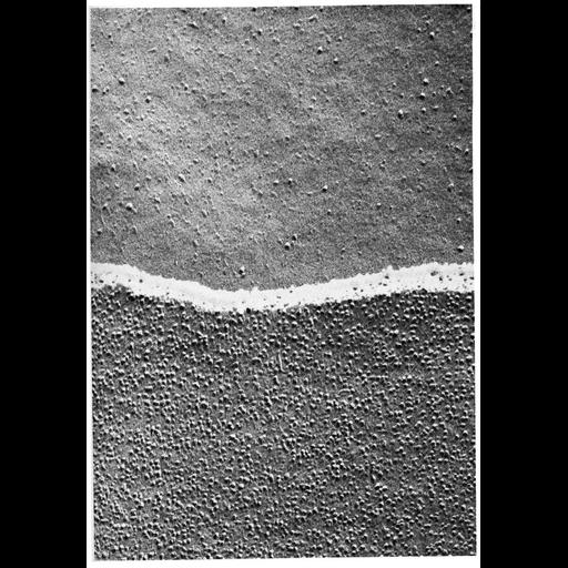
This electron micrograph shows the E-fracture face (above) and P-fracture face (below) of the membrane of adjacent epithelial cells from the ciliary epithelium of the monkey eye. Intramembranous particles, representing integral membrane proteins, appear as bumpy projections, and are prominent in this image with high density on the p-face, but also can be detected on the e-face. This image, contributed by Giuseppina Raviola, is Figure 4 from Chapter 1 (The Cell Surface) of 'The Cell, 2nd Ed.' by Don W. Fawcett M.D. A PDF copy of the accompanying chapter is available on the ASCB's BioEDUCATE website.
With the freeze-fracture technique, tissue is rapidly frozen and cracked to shear along zones of weakness. Cleavage of membranes occurs along the hydrophobic interior of the lipid bilayer to reveal views of a "p-face" (the outwardly-facing inner half of the membrane) and an "e-face" (the inwardly-facing outer half of the membrane), and a metallic replica is made of the fractured surface.
| Spatial Axis | Image Size | Pixel Size |
|---|---|---|
| X | 898px | —— |
| Y | 1295px | —— |