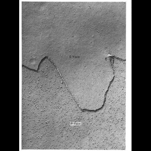
Freeze-fracture preparation of the plasma membrane from an unspecialized area composed of Sertoli cells of the guinea pig testis. With the freeze-fracture technique, tissue is rapidly frozen and cracked to shear along zones of weakness. Cleavage of membranes occurs along the hydrophobic interior of the lipid bilayer to reveal views of a "p-face" (the outwardly-facing inner half of the membrane) and an "e-face" (the inwardly-facing outer half of the membrane), and a metallic replica is made of the fractured surface. Intramembranous particles, representing integral membrane proteins, appear as bumpy projections, and are prominent on the p-face, but also can be detected on the e-face. The particles on the p-face range in size from 6 - 9 nm. Figure 3 from Chapter 1 (The Cell Surface) of 'The Cell, 2nd Ed.' by Don W. Fawcett M.D. A PDF copy of the accompanying chapter is available on the ASCB's BioEDUCATE website.
| Spatial Axis | Image Size | Pixel Size |
|---|---|---|
| X | 964px | —— |
| Y | 1294px | —— |