Alternate header for print version


Licensing
Attribution Non-Commercial; No Derivatives:This image is licensed under a Creative Commons Attribution, Non-Commercial, No Derivatives License. View License Deed | View Legal Code
tweet  
*CIL – Cell Image Library accession number. Please use this to reference an image.

CIL:10840*  Cite 
Description

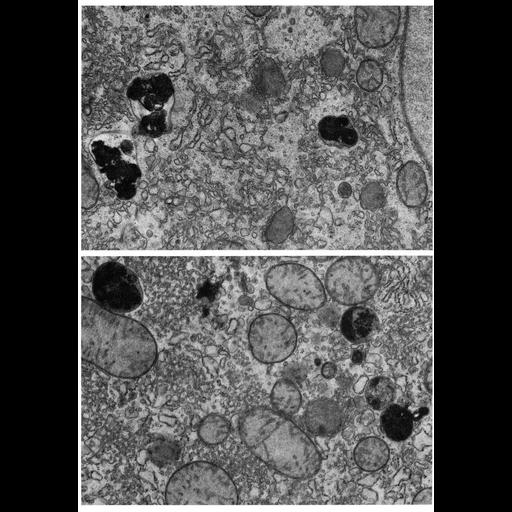
Figures 264 (upper) and 265 (lower) from Chapter 8 (Lysosomes) of 'The Cell, 2nd Ed.' by Don W. Fawcett M.D. Lysosomes apparent in thin sections from liver tissue of hamster treated with phenobarbitol. Primary lysosomes have dense homogeneous content, but the majority of the membrane-bounded dense bodies in the accompanying figures are secondary lysosomes. A PDF copy of the accompanying chapter is available on the ASCB’s BioEDUCATE website.

Biological Sources
NCBI Organism Classification
Phodopus
Cell Type
hepatocyte
Cellular Component
lysosome
primary lysosome
secondary lysosome
Biological Context
Biological Process
autophagy
Attribution
Names
Don W. Fawcett
Published
ISBN 0721635849
Link
http://www.ascb.org/bioeducate/FawcettTheCell...
Citation
Digital Object Identifier (DOI)
doi:10.7295/W9CIL10840
Archival Resource Key (ARK)
ark:/b7295/w9cil10840
Grouping This image is part of a group.
Imaging
Image Type
transmission electron microscopy (TEM)
Image Mode
illumination by electrons
detection of electrons
Parameters Imaged
electron density
Source of Contrast
stain with broad specificity
Data Qualifiers
processed data
Dimensions
Spatial Axis Image Size Pixel Size
X 908px ——
Y 1304px ——