Alternate header for print version


Licensing
Attribution Non-Commercial; No Derivatives:This image is licensed under a Creative Commons Attribution, Non-Commercial, No Derivatives License. View License Deed | View Legal Code
tweet  
*CIL – Cell Image Library accession number. Please use this to reference an image.

CIL:10788*  Cite 
Description

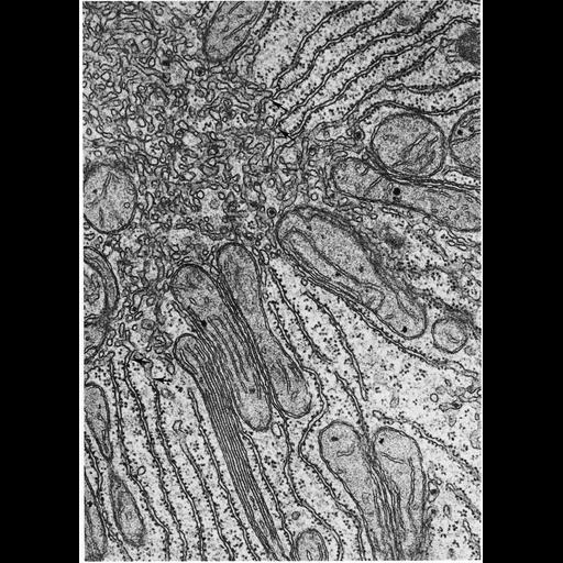
Figure 179 from Chapter 5 (Endoplasmic Reticulum) of 'The Cell, 2nd Ed.' by Don W. Fawcett M.D. Electron micrograph showing a portion of the cytoplasm of a liver cell. The upper left part of the micrograph shows a network of branching and anastomosing smooth tubules that appear to connect with the cisternae of the rough endoplasmic reticulum (points of interconnection indicated by arrows). Image by Robert Bolander. A PDF copy of the accompanying chapter is available on the ASCB’s BioEDUCATE website.

Biological Context
Biological Process
translation
Attribution
Names
Robert Bolender
Published
ISBN 0721635849
Link
http://www.ascb.org/bioeducate/FawcettTheCell...
Citation
Digital Object Identifier (DOI)
doi:10.7295/W9CIL10788
Archival Resource Key (ARK)
ark:/b7295/w9cil10788
Grouping This image is part of a group.
Imaging
Image Type
transmission electron microscopy (TEM)
Image Mode
illumination by electrons
detection of electrons
Parameters Imaged
electron density
Source of Contrast
stain with broad specificity
Data Qualifiers
processed data
Dimensions
Spatial Axis Image Size Pixel Size
X 906px ——
Y 1264px ——