Alternate header for print version


Licensing
Attribution Non-Commercial; No Derivatives:This image is licensed under a Creative Commons Attribution, Non-Commercial, No Derivatives License. View License Deed | View Legal Code
tweet  
*CIL – Cell Image Library accession number. Please use this to reference an image.

CIL:10769*  Cite 
Description

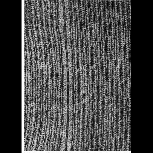
Figure 171 from Chapter 5 (Endoplasmic Reticulum) of 'The Cell, 2nd Ed.' by Don W. Fawcett M.D. Electron micrograph of rough endoplasmic reticulum (RER) in two adjacent acinar cells from the pancreas of the small brown bat, Myotis lucifugus. The surface membranes for the two cells are closely apposed and slightly thicker than the membranes associated with the ER. While free ribosomes are apparent between the plasma membrane and the ER, the ribosomes are separated from the inner border of the plasma membrane by about 10 nm (arrows). In contrast, the membrane associated with the ER is studded with adherent ribosomes. A PDF copy of the accompanying chapter is available on the ASCB’s BioEDUCATE website.

Biological Sources
NCBI Organism Classification
Myotis lucifugus
Cell Type
acinar cell
Cellular Component
endoplasmic reticulum
ribosome
rough endoplasmic reticulum
endoplasmic reticulum cisternal network
Biological Context
Biological Process
translation
Attribution
Names
Don W. Fawcett
Published
ISBN 0721635849
Link
http://www.ascb.org/bioeducate/FawcettTheCell...
Citation
Digital Object Identifier (DOI)
doi:10.7295/W9CIL10769
Archival Resource Key (ARK)
ark:/b7295/w9cil10769
Grouping This image is part of a group.
Imaging
Image Type
transmission electron microscopy (TEM)
Image Mode
illumination by electrons
detection of electrons
Parameters Imaged
electron density
Source of Contrast
stain with broad specificity
Data Qualifiers
processed data
Dimensions
Spatial Axis Image Size Pixel Size
X 920px ——
Y 1280px ——