Alternate header for print version


Licensing
Attribution Non-Commercial; No Derivatives:This image is licensed under a Creative Commons Attribution, Non-Commercial, No Derivatives License. View License Deed | View Legal Code
tweet  
*CIL – Cell Image Library accession number. Please use this to reference an image.

CIL:10763*  Cite 
Description

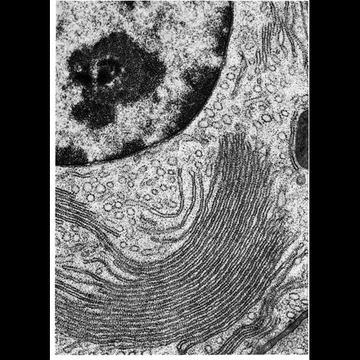
Figure 168 from Chapter 5 (Endoplasmic Reticulum) of 'The Cell, 2nd Ed.' by Don W. Fawcett M.D. Electron micrograph of rough endoplasmic reticulum (RER) in an acinar cell from the pancreas of the small brown bat, Myotis lucifugus. The nucleus of the cell is in the upper left corner; the RER in the lower half of the micrograph is stacked in a cisternal structure, and studded with ribosomes. This cisternal packed organization is common in cells that actively produce proteins for secretion, like this acinar cell. A PDF copy of the accompanying chapter is available on the ASCB’s BioEDUCATE website.

Biological Context
Biological Process
translation
Attribution
Names
Don W. Fawcett
Published
ISBN 0721635849
Link
http://www.ascb.org/bioeducate/FawcettTheCell...
Citation
Digital Object Identifier (DOI)
doi:10.7295/W9CIL10763
Archival Resource Key (ARK)
ark:/b7295/w9cil10763
Grouping This image is part of a group.
Imaging
Image Type
transmission electron microscopy (TEM)
Image Mode
illumination by electrons
detection of electrons
Parameters Imaged
electron density
Source of Contrast
stain with broad specificity
Data Qualifiers
processed data
Dimensions
Spatial Axis Image Size Pixel Size
X 918px ——
Y 1272px ——