Alternate header for print version


Licensing
Public Domain: This image is in the public domain and thus free of any copyright restrictions. However, as is the norm in scientific publishing and as a matter of courtesy, any user should credit the content provider for any public or private use of this image whenever possible. Learn more
tweet  
*CIL – Cell Image Library accession number. Please use this to reference an image.

CIL:10488*  Cite 
Description

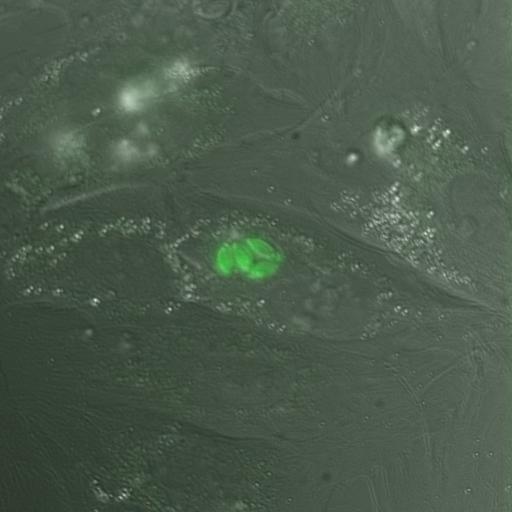
A monolayer of human foreskin fibroblast cells was infected with Toxoplasma gondii expressing EGFP-tagged Tgbeta3-tubulin. 50 timepoints in which daughter formation is visible. Images overlaid with DIC. Images acquired using API DeltaVision on an Olympus IX70 inverted microscope. Objective: Olympus 60X water immersion, 1.2 NA. Filters: GFP, ex 450-490 em 500-540. Captured with CoolSnapHQ/ICX285. 3D stack is raw data corresponding to deconvolved stack: CIL# 10489. Unpublished image that is similar to images published in PMID: 16518471. See publication for other experimental details.

Biological Sources
NCBI Organism Classification
Toxoplasma gondii RH
Cell Line
Toxoplasma gondii RH stable cell line
Cellular Component
cortical microtubule
Attribution
Names
John Murray (University of Pennsylvania)
Citation
Digital Object Identifier (DOI)
doi:10.7295/W9CIL10488
Archival Resource Key (ARK)
ark:/b7295/w9cil10488
Grouping This image is part of a group.
Sample Preparation
Methods
living tissue
Relation To Intact Cell
whole mounted tissue
Dimensions
Spatial Axis Image Size Pixel Size
X 1024px 0.1067µm
Y 1024px 0.1067µm
Z 8px 0.5µm
Time 304 seconds 50