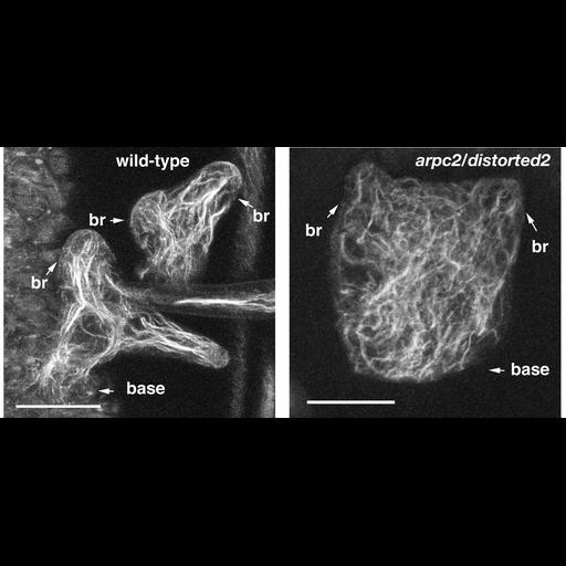
Polymerized actin localization in wild type (left) and mutant (right) Arabidopsis thaliana leaf trichomes during the transition to branch elongation. The "distorted group" of trichome morphology mutants were originally identified as a class of mutants that failed to maintain polarized growth following branch initiation. Subsequent analyses indicated that distorted mutants are specifically affected in actin-dependent morphogenesis and fail to organize a polarized cytoskeleton in developing branches. Genetic experiments later proved that DISTORTED genes encode subunits of the evolutionarily conserved WAVE and ARP2/3 complexes. These heteromeric complexes translate Rho family small GTPase signals into actin filament nucleation and a coordinated growth response. Detailed methods: Intact Arabidopsis shoots were fixed in 100 mM PIPES/KOH pH 6.9, 5 mM EGTA, 4 mM MgCl2 (PEM) containing 2 % formaldehyde. Actin filaments were detected with overnight incubation with 0.2 µM Alexa Fluor 488® phalloidin. Confocal images were obtains using a Biorad Radiance 2100 confocal head mounted on a Nikon E800 stand. The objective used was a 60X 1.2 NA water immersion lens. Acquisition Parameters: Still image. Maximum projections of confocal images of the actin cytoskeleton in normal (left panel) and the ARP2/3 complex subunit mutant arpc2/distorted2 (right). Trichomes are unicellular structures that arise from single protodermal cells and usually form three highly elongated branches. br=branch, base=attachment region of the cell. Magnification: bar=20 µm. For more information see these references: Hulskamp, M., Misra, S., Jurgens, G. Genetic dissection of trichome cell development in Arabidopsis, Cell 1994, 76: 555-566. Szymanski, D. Breaking the WAVE complex: the point of Arabidopsis trichomes. Curr Opin Plant Biol 2005, 8: 103-112. The maximum projection image from the left panel has appeared as part of Fig. 3 in Kotchoni et al. (2009) Plant Physio. 151: 2095-109. Copyright American Society of Plant Biologists
| Spatial Axis | Image Size | Pixel Size |
|---|---|---|
| X | 2500px | 0.0526µm |
| Y | 1200px | 0.0526µm |