Alternate header for print version


Licensing
This image is copyright protected. Any public or private use of this image is subject to prevailing copyright laws. Please contact the content provider of this image for permission requests.
tweet  
*CIL – Cell Image Library accession number. Please use this to reference an image.

CIL:10276*  Cite 
Description

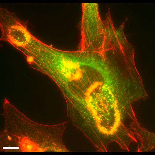
After over-night serum starvation, mouse fibroblast expressing Dyn2-GFP were stimulated with 10ng/ml PDGF. After 5min, the cells were fixed and stained with rhodamine-phalloidin. PDGF-induced dorsal membrane ruffle was indicated by F-actin staining, and the enrichment of dynamin to the membrane ruffle was shown by their co-localization. Biological Source Mus musculus ES cell derived fibroblast PDGF-induced macropinocytosis PFA fixation Biological Context and Probe PDGF-induced dorsal membrane ruffle, Dyn2-GFP and rhodamine-phalloidin Equipment used Inverted Olympus IX-70 microscope with a 100×, 1.35 NA oil-immersion objective Magnification of image 1000x, scale bar indicates 5 um

Biological Sources
NCBI Organism Classification
Mus musculus
Cell Type
fibroblast
Cellular Component
dynamin
actin cytoskeleton
dorsal membrane ruffle
Attribution
Names
Ya-Wen Li
Sandra L. Schmid
Citation
Digital Object Identifier (DOI)
doi:10.7295/W9CIL10276
Archival Resource Key (ARK)
ark:/b7295/w9cil10276
Imaging
Image Type
recorded image
Image Mode
fluorescence microscopy
Parameters Imaged
fluorescence emission
Source of Contrast
distribution of a specific protein
Visualization Methods
phalloidin
Rhodamine
EGFP
Processing History
unprocessed raw data
Sample Preparation
Relation To Intact Cell
dispersed cells in vitro
Dimensions
Spatial Axis Image Size Pixel Size
X 801px 0.059µm
Y 770px 0.059µm
Channel Wavelength
1 GFP, Rhodamineµm