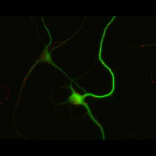
Early stages of dendritic development and synapse formation in cultured hippocampal neurons. This multilayer image shows neurons fixed at 7 days in vitro and immunostained for the dendritically localized protein MAP2 (green) and the presynaptic vesicle protein Synapsin I (red). Unstained axons in the field are evident in the phase channel, which is hidden but can be turned on in the viewer. Neurons at 3, 5 and 7 days in vitro are represented in this image group. Detailed methods: Embryonic rat hippocampal neurons were prepared as previously described (see Kaech and Banker, 2006, Nat Protoc). Cells were prepared for fluorescent staining as previously described (Withers and Banker, 1998, in Culturing Nerve Cells, MIT Press). Briefly, cells were fixed (4% formaldehyde, 4% sucrose in phosphate buffered saline, pH 7.4, warmed to 37°C prior to fixation, 15 minutes), permeabilized with 0.25% Triton (7 minutes) and immunostained for MAP2 (monoclonal HM2, Sigma, with Alexa 488 conjugated secondary, excitation, 494, emission, 519 [Invitrogen, Molecular Probes]) and synapsin I (from P. DeCamilli, with DyLight549 conjugated secondary, excitation, 555, emission, 568, [Jackson Immunoresearch]). Images were acquired with a Leica DMRA microscope with a mercury arc lamp, a 40X lens (HCX PL Fluotar, NA 0.75), Leica GFP filter set (excitation, BP 470/40; dichromatic mirror, 500; suppression filter, BP 525/50); Leica N3 filter set (excitation, BP546/12; dichromatic mirror, 565, suppression filter, BP 600/40), Photometrics CoolSnap ES CCD camera and MetaMorph software.
| Spatial Axis | Image Size | Pixel Size |
|---|---|---|
| X | 1300px | 0.167µm |
| Y | 1030px | 0.167µm |