Alternate header for print version


Licensing
Attribution Non-Commercial Share Alike:This image is licensed under a Creative Commons Attribution, Non-Commercial Share Alike License. View License Deed | View Legal Code
tweet  
*CIL – Cell Image Library accession number. Please use this to reference an image.

CIL:10093*  Cite 
Description

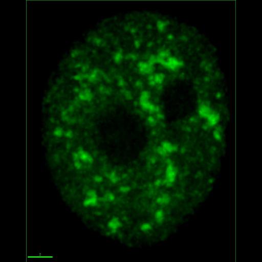
Projection of a HeLa cell expressing a YFP-SC35 fusion protein that localizes to nuclear speckles and in a diffuse nuclear localization. Image stack was acquired using a Deltavision deconvolution microscope.

Biological Sources
NCBI Organism Classification
Homo sapiens
Cell Type
cervical carcinoma
Cell Line
HeLa
Cellular Component
nucleus
Biological Context
Biological Process
RNA splicing
RNA metabolic process
Attribution
Names
R.I. Kumaran
Z. Lazar
David L. Spector
Citation
Digital Object Identifier (DOI)
doi:10.7295/W9CIL10093
Archival Resource Key (ARK)
ark:/b7295/w9cil10093
Imaging
Image Type
recorded image
Image Mode
fluorescence microscopy
constrained iterative deconvolution
charge coupled device (CCD)
Parameters Imaged
fluorescence emission
Source of Contrast
level of fluorochrome
Visualization Methods
EYFP
Processing History
constrained iterative deconvolution
projection of image stack
Application of green LUT
Sample Preparation
Methods
living tissue
Relation To Intact Cell
whole mounted tissue
Dimensions
Spatial Axis Image Size Pixel Size
X 857px 40nm
Y 1071px 40nm