Licensing

Pd
Public Domain: This image is in the public domain and thus free of any copyright restrictions. However, as is the norm in scientific publishing and as a matter of courtesy, any user should credit the content provider for any public or private use of this image whenever possible. Learn more
Comments: Powered by Pnyxe

Description

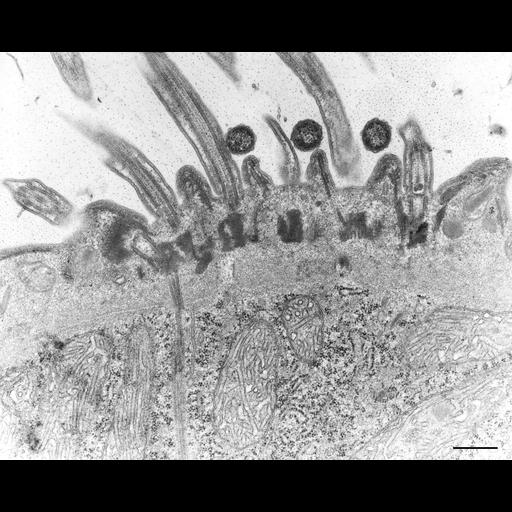
The locomotor cortex of Didinium. A longitudinal section of basal bodies and the axonemes of cilia is revealed as they emerge from the cell cortex of Didinium. Bundles of microtubules extend from the basal bodies into the endoplasm through the fibrous layer that characterizes the cortex of Didinium. Tubular or flattened early endosomes with coated pits at their margins are present in the endoplasm beneath the fibrous layer. Mitochondrial cristae appear to be decorated by F1F0 ATP synthases. Cross sections of three bacteria are noted between the cilia. TEM taken on 5/9/69 by M. Sage with Philips 300 operating at 60kV. Neg. 14,800X. Bar = 0.5µm. The negative was printed to paper and the image was scanned to Photoshop. This digitized image is available for qualitative analysis. A raw, unprocessed, high resolution version of this image (CIL:4661) is in the library and available for quantitative analysis. Standard glutaraldehyde fixation followed by osmium tetroxide, dehydrated in alcohol and embedded in an epoxy resin. Microtome sections prepared at approximately 75nm thickness. Additional information available at (http://www5.pbrc.hawaii.edu/allen/).

Biological Sources

NCBI Organism Classification
Didinium nasutum
Cell Type
eukaryotic cell
Eukaryotic Protist
Ciliated Protist
Cell Line
Carolina Biological Supply Company, NC, U.S.A.
Cellular Component
cell cortex
cilium
microtubule basal body
ciliary rootlet
mitochondrion

Biological Context

Biological Process
ciliary cell motility
microtubule cytoskeleton organization
Molecular Function
cytoskeletal protein binding

Attribution

Names
M. Sage
Richard Allen
Link
http://www5.pbrc.hawaii.edu/allen/

Grouping

This image is part of a group.

Imaging

Image Type
transmission electron microscopy (TEM)
illumination by electrons
Imaging Mode
detection of electrons
film
Parameters Imaged
electron density
Source of Contrast
stain with broad specificity
Visualization Methods
stain with broad specificity
osmium tetroxide
uranyl salt
lead salt
Processing History
film
Print from negative scanned for Photoshop.
Data Qualifiers
processed data
suitable for spatial measurements

Dimensions

Spatial Axis Image Size Pixel Size
X 3496px ——
Y 2792px ——
*CIL – Cell Image Library accession number. Please use this to reference an image.