Alternate header for print version


Licensing
Public Domain: This image is in the public domain and thus free of any copyright restrictions. However, as is the norm in scientific publishing and as a matter of courtesy, any user should credit the content provider for any public or private use of this image whenever possible. Learn more
tweet  
*CIL – Cell Image Library accession number. Please use this to reference an image.

CIL:10008*  Cite 
Description

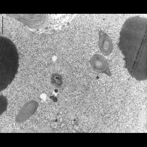
Toxicysts in the cytoplasm of cross sectioned non-dividing Didinium. Toxicysts (extrusomes) are multilayered cylinders that are discharged from the cytopharynx region during capture of prey such as Paramecium. Lipid is stored as large non-membrane enclosed dense bodies in the cytosol. TEM taken on 2/18/69 by R. Allen with Philips 300. Neg. 20,500X. Bar = 0.5µm. The negative was printed to paper and the image was scanned to Photoshop. This digitized image is available for qualitative analysis. A raw, unprocessed, high resolution version of this image (CIL:9929) is in the library and available for quantitative analysis. Standard glutaraldehyde fixation followed by osmium tetroxide, dehydrated in alcohol and embedded in an epoxy resin. Microtome sections prepared at approximately 75nm thickness. Additional information available at (http://www5.pbrc.hawaii.edu/allen/).

Biological Sources
NCBI Organism Classification
Didinium nasutum
Cell Type
eukaryotic cell
Eukaryotic Protist
Ciliated Protist
Cell Line
Carolina Biological Supply Company, NC, U.S.A.
Cellular Component
rough endoplasmic reticulum
lipid particle
extrusome
toxicyst
Biological Context
Biological Process
cytoplasm organization
organelle organization
organelle localization
organelle development
Attribution
Names
Richard Allen
Link
http://www5.pbrc.hawaii.edu/allen/
Citation
Digital Object Identifier (DOI)
doi:10.7295/W9CIL10008
Archival Resource Key (ARK)
ark:/b7295/w9cil10008
Grouping This image is part of a group.
Imaging
Image Type
transmission electron microscopy (TEM)
illumination by electrons
Image Mode
detection of electrons
film
Parameters Imaged
electron density
Source of Contrast
stain with broad specificity
Visualization Methods
stain with broad specificity
osmium tetroxide
uranyl salt
lead salt
Processing History
film
Print from negative scanned for Photoshop.
Dimensions
Spatial Axis Image Size Pixel Size
X 3732px ——
Y 3032px ——