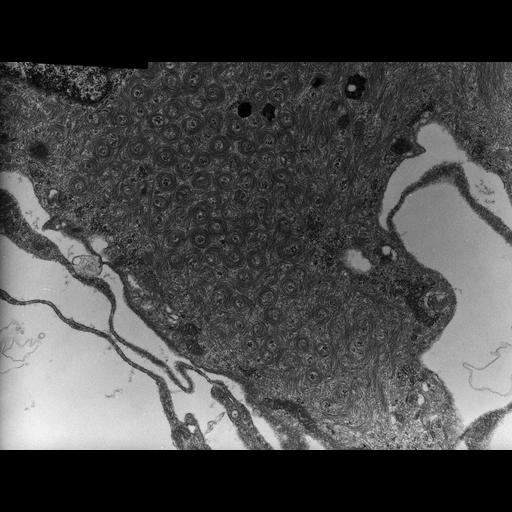
This group of micrographs illustrate the ultrastructural characteristics of the middle layer of the three meningeal layers that cover the central nervous system The arachnoid cells contain abundant intermediate filaments that occupy a substantial volume of the cytoplasm. In some cells the intermediate filaments are organized into numerous coils of relatively uniform size and orientation. The coils appear to stack in loose hexagonal arrays. The plasma membrane of these cells has many caveolae. Adjacent arachnoid cells are connected by large desmosomes. Images were recorded using a Philips EM301 TEM at 80 kv. Original glass plates were scanned and digitized using an Epson V750 flatbed scanner with a step size of 15 micrometers. This group of images complement those published in the reference below. Reference: Michaels, JE, Tornheim, PA. Cell Tissue Res. 236:693-697 (1984).
| Spatial Axis | Image Size | Pixel Size |
|---|---|---|
| X | 6103px | 1.32nm |
| Y | 4628px | 1.32nm |