Alternate header for print version
Advanced search
Contributors
Help
Submit
Search
menu
Cell Process
Cell Component
Cell Type
Organism
Microbial
Alzheimer's
Data Sets
University of California, San Diego
9500 Gilman Drive
La Jolla, CA 92093-0608, USA
Voice
: (858) 534-0276
Fax
: (858) 534-7497
Email
: dorloff@ncmir.ucsd.edu
Grouped images - the images shown below are related
CIL:24783
NCBI Organism Classification
Xenopus laevis
Biological Process
cellular localization
Cellular Component
lamellipodium
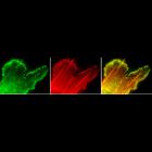
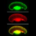
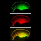
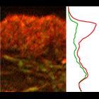
Localization of XAC (Xenopus ADF/cofilin) in Xenopus fibroblasts. Fluorescence m...
CIL:24784
NCBI Organism Classification
Xenopus laevis
Biological Process
actin filament organization
Cellular Component
lamellipodium
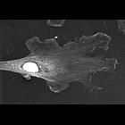
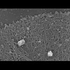
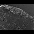
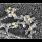
Localization of XAC (Xenopus ADF/cofilin) in Xenopus fibroblasts. Immuno-EM with...
CIL:24785
NCBI Organism Classification
Xenopus laevis
Biological Process
actin filament-based process
Cellular Component
lamellipodium
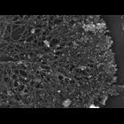
Localization of XAC (Xenopus ADF/cofilin) in Xenopus fibroblasts. Immuno-EM with...
CIL:24786
NCBI Organism Classification
Xenopus laevis
Biological Process
branching of actin filaments
Cellular Component
lamellipodium
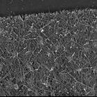
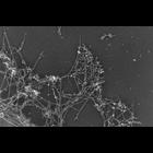
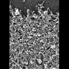
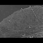
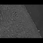
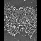
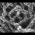
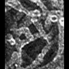
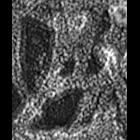
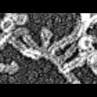
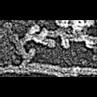
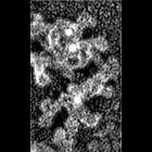
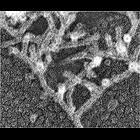
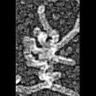
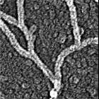
Multiple branching of actin filaments in lamellipodia of Xenopus keratocytes. Th...
CIL:24788
NCBI Organism Classification
none specified
Biological Process
branching of actin filaments
Cellular Component
lamellipodium
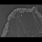
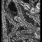
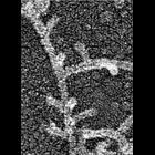
Multiple branching of actin filaments in lamellipodia of vertebrate fibroblasts....
CIL:24790
NCBI Organism Classification
none specified
Biological Process
branching of actin filaments
Cellular Component
lamellipodium
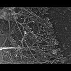
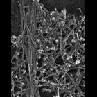
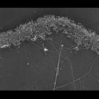
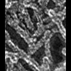
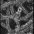
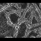
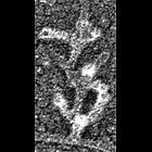
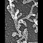
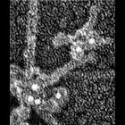
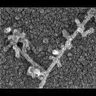
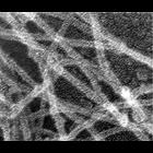
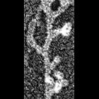
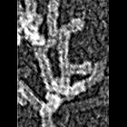
Improved visualization of actin filament branching in lamellipodia. EM of kerato...
CIL:24792
NCBI Organism Classification
none specified
Biological Process
branching of actin filaments
Cellular Component
actin cytoskeleton
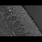
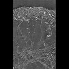
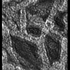
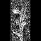
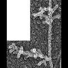
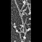
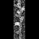
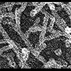
Electron micrograph of keratocyte or fibroblast lamellipodial actin network afte...
CIL:24794
NCBI Organism Classification
Xenopus laevis
Biological Process
cellular macromolecule localization
Cellular Component
lamellipodium
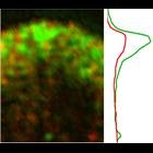
Localization of Arp2/3 complex in the lamellioidia of a Xenopus keratocytes. Sta...
CIL:24795
NCBI Organism Classification
Xenopus laevis
Biological Process
cellular macromolecule localization
Cellular Component
lamellipodium
Localization of Arp2/3 complex in the lamellioidia of a Xenopus keratocytes. Sta...
CIL:24796
NCBI Organism Classification
none specified
Biological Process
cellular macromolecule localization
Cellular Component
lamellipodium
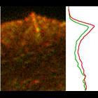
Localization of Arp2/3 complex in a fibroblast lamellioidia. Staining with p21 a...
CIL:24797
NCBI Organism Classification
none specified
Biological Process
actin filament-based process
Cellular Component
lamellipodium
Localization of Arp2/3 complex in the lamellioidia of afibroblast . Staining wit...
CIL:24800
NCBI Organism Classification
Xenopus laevis
Biological Process
actin filament organization
Cellular Component
lamellipodium
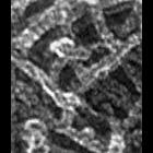
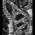
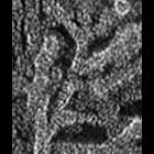
Structural differentiation of actin network in lamellipodium. Electron micrograp...
CIL:24801
NCBI Organism Classification
Xenopus laevis
Biological Process
cellular localization
Cellular Component
lamellipodium
Structural differentiation of actin network in lamellipodium. Electron micrograp...
CIL:24802
NCBI Organism Classification
Xenopus laevis
Biological Process
cellular localization
Cellular Component
lamellipodium
Structural differentiation of actin network in lamellipodium. Electon micrograph...
CIL:24803
NCBI Organism Classification
Xenopus laevis
Biological Process
cellular localization
Cellular Component
lamellipodium
Structural differentiation of actin network in lamellipodium. Electron micrograp...
CIL:24804
NCBI Organism Classification
Xenopus laevis
Biological Process
branching of actin filaments
Cellular Component
actin cytoskeleton
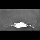
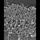
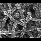
Differential response of lamellipodial actin network to latrunculin a (LA), a bi...
CIL:24805
NCBI Organism Classification
Xenopus laevis
Biological Process
branching of actin filaments
Cellular Component
actin cytoskeleton
Differential response of lamellipodial actin network to latrunculin a (LA). Elec...
CIL:24806
NCBI Organism Classification
Xenopus laevis
Biological Process
cellular localization
Cellular Component
lamellipodium
Localization of XAC (Xenopus ADF/cofilin) in Xenopus keratocytes. Fluorescence m...
CIL:24807
NCBI Organism Classification
Xenopus laevis
Biological Process
cellular localization
Cellular Component
lamellipodium
Localization of XAC (Xenopus ADF/cofilin) in Xenopus keratocytes done with immun...
CIL:24808
NCBI Organism Classification
Xenopus laevis
Biological Process
cellular localization
Cellular Component
lamellipodium
Localization of XAC (Xenopus ADF/cofilin) in Xenopus keratocytes done with immun...
CIL:24809
NCBI Organism Classification
Xenopus laevis
Biological Process
cellular localization
Cellular Component
lamellipodium
Localization of Xenopus ADF/cofilin (XAC) to posterior regions of depolymerizati...
CIL:24810
NCBI Organism Classification
Xenopus laevis
Biological Process
latrunculin a treatment
Cellular Component
lamellipodium
Localization of XAC XAC (Xenopus ADF/cofilin) to posterior regions of depolymeri...
CIL:24811
NCBI Organism Classification
Xenopus laevis
Biological Process
cellular localization
Cellular Component
lamellipodium
Localization of Xenopus ADF/cofilin (XAC) to posterior regions of depolymerizati...
CIL:34882
NCBI Organism Classification
Xenopus laevis
Biological Process
branching of actin filaments
Cellular Component
actin cytoskeleton
Multiple branching of actin filaments in lamellipodia of Xenopus keratocytes. Th...
CIL:34883
NCBI Organism Classification
Xenopus laevis
Biological Process
branching of actin filaments
Cellular Component
actin cytoskeleton
Multiple branching of actin filaments in lamellipodia of Xenopus keratocytes. Th...
CIL:34884
NCBI Organism Classification
Xenopus laevis
Biological Process
branching of actin filaments
Cellular Component
actin cytoskeleton
Multiple branching of actin filaments in lamellipodia of Xenopus keratocytes. Th...
CIL:34885
NCBI Organism Classification
Xenopus laevis
Biological Process
branching of actin filaments
Cellular Component
actin cytoskeleton
Multiple branching of actin filaments in lamellipodia of Xenopus keratocytes. Th...
CIL:34886
NCBI Organism Classification
Xenopus laevis
Biological Process
branching of actin filaments
Cellular Component
actin cytoskeleton
Multiple branching of actin filaments in lamellipodia of Xenopus keratocytes. Th...
CIL:34887
NCBI Organism Classification
Xenopus laevis
Biological Process
branching of actin filaments
Cellular Component
actin cytoskeleton
Multiple branching of actin filaments in lamellipodia of Xenopus keratocytes. Th...
CIL:34888
NCBI Organism Classification
none specified
Biological Process
branching of actin filaments
Cellular Component
actin cytoskeleton
Multiple branching of actin filaments in lamellipodia of vertebrate fibroblasts....
CIL:34889
NCBI Organism Classification
none specified
Biological Process
branching of actin filaments
Cellular Component
actin cytoskeleton
Multiple branching of actin filaments in lamellipodia of vertebrate fibroblasts....
CIL:34890
NCBI Organism Classification
none specified
Biological Process
branching of actin filaments
Cellular Component
actin cytoskeleton
Multiple branching of actin filaments in lamellipodia of vertebrate fibroblasts....
CIL:34891
NCBI Organism Classification
none specified
Biological Process
branching of actin filaments
Cellular Component
actin cytoskeleton
Multiple branching of actin filaments in lamellipodia of vertebrate fibroblasts....
CIL:34892
NCBI Organism Classification
none specified
Biological Process
branching of actin filaments
Cellular Component
actin cytoskeleton
Multiple branching of actin filaments in lamellipodia of vertebrate fibroblasts....
CIL:34893
NCBI Organism Classification
none specified
Biological Process
branching of actin filaments
Cellular Component
actin cytoskeleton
Multiple branching of actin filaments in lamellipodia of vertebrate fibroblasts....
CIL:34894
NCBI Organism Classification
none specified
Biological Process
branching of actin filaments
Cellular Component
actin cytoskeleton
Improved visualization of actin filament branching in lamellipodia. EM of kerato...
CIL:34895
NCBI Organism Classification
none specified
Biological Process
branching of actin filaments
Cellular Component
actin cytoskeleton
Improved visualization of actin filament branching in lamellipodia. EM of kerato...
CIL:34896
NCBI Organism Classification
none specified
Biological Process
branching of actin filaments
Cellular Component
actin cytoskeleton
Improved visualization of actin filament branching in lamellipodia. EM of kerato...
CIL:34897
NCBI Organism Classification
none specified
Biological Process
branching of actin filaments
Cellular Component
actin cytoskeleton
Improved visualization of actin filament branching in lamellipodia. EM of kerato...
CIL:34898
NCBI Organism Classification
none specified
Biological Process
branching of actin filaments
Cellular Component
actin cytoskeleton
Improved visualization of actin filament branching in lamellipodia. EM of kerato...
CIL:34899
NCBI Organism Classification
none specified
Biological Process
branching of actin filaments
Cellular Component
actin cytoskeleton
Improved visualization of actin filament branching in lamellipodia. EM of kerat...
CIL:34900
NCBI Organism Classification
none specified
Biological Process
branching of actin filaments
Cellular Component
actin cytoskeleton
Improved visualization of actin filament branching in lamellipodia. EM of kerato...
CIL:34901
NCBI Organism Classification
none specified
Biological Process
branching of actin filaments
Cellular Component
actin cytoskeleton
Improved visualization of actin filament branching in lamellipodia. EM of kerato...
CIL:34902
NCBI Organism Classification
none specified
Biological Process
branching of actin filaments
Cellular Component
actin cytoskeleton
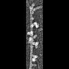
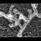
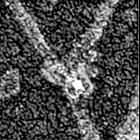
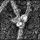
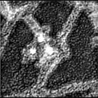
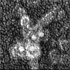
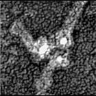
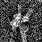
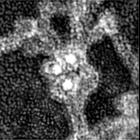
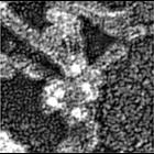
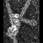
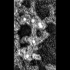
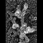
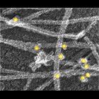
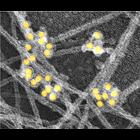
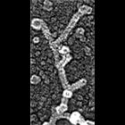
Localization of Arp2/3 complex at actin filament branching points. Xenopus kerat...
CIL:34903
NCBI Organism Classification
none specified
Biological Process
branching of actin filaments
Cellular Component
actin cytoskeleton
Localization of Arp2/3 complex at actin filament branching points. Xenopus kerat...
CIL:34904
NCBI Organism Classification
none specified
Biological Process
branching of actin filaments
Cellular Component
actin cytoskeleton
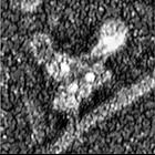
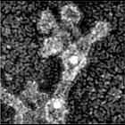
Localization of Arp2/3 complex at actin filament branching points. Xenopus kerat...
CIL:34905
NCBI Organism Classification
none specified
Biological Process
branching of actin filaments
Cellular Component
actin cytoskeleton
Localization of Arp2/3 complex at actin filament branching points. Xenopus kerat...
CIL:34906
NCBI Organism Classification
none specified
Biological Process
branching of actin filaments
Cellular Component
actin cytoskeleton
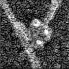
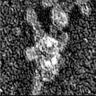
Localization of Arp2/3 complex at actin filament branching points. Xenopus kerat...
CIL:34907
NCBI Organism Classification
none specified
Biological Process
branching of actin filaments
Cellular Component
actin cytoskeleton
Localization of Arp2/3 complex at actin filament branching points. Xenopus kerat...
CIL:34908
NCBI Organism Classification
none specified
Biological Process
branching of actin filaments
Cellular Component
actin cytoskeleton
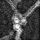
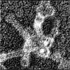
Localization of Arp2/3 complex at actin filament branching points. Xenopus kerat...
CIL:34909
NCBI Organism Classification
none specified
Biological Process
branching of actin filaments
Cellular Component
actin cytoskeleton
Localization of Arp2/3 complex at actin filament branching points. Xenopus kerat...
CIL:34910
NCBI Organism Classification
none specified
Biological Process
branching of actin filaments
Cellular Component
actin cytoskeleton
Localization of Arp2/3 complex at actin filament branching points. Xenopus kerat...
CIL:34911
NCBI Organism Classification
none specified
Biological Process
branching of actin filaments
Cellular Component
actin cytoskeleton
Localization of Arp2/3 complex at actin filament branching points. Xenopus kerat...
CIL:34912
NCBI Organism Classification
none specified
Biological Process
branching of actin filaments
Cellular Component
actin cytoskeleton
Localization of Arp2/3 complex at actin filament branching points. Xenopus kerat...
CIL:34913
NCBI Organism Classification
none specified
Biological Process
branching of actin filaments
Cellular Component
actin cytoskeleton
Localization of Arp2/3 complex at actin filament branching points. Xenopus kerat...
CIL:34914
NCBI Organism Classification
none specified
Biological Process
branching of actin filaments
Cellular Component
actin cytoskeleton
Localization of Arp2/3 complex at actin filament branching points. Xenopus kerat...
CIL:34915
NCBI Organism Classification
none specified
Biological Process
branching of actin filaments
Cellular Component
actin cytoskeleton
Localization of Arp2/3 complex at actin filament branching points. Xenopus kerat...
CIL:34916
NCBI Organism Classification
none specified
Biological Process
branching of actin filaments
Cellular Component
actin cytoskeleton
Localization of Arp2/3 complex at actin filament branching points. Xenopus kerat...
CIL:34917
NCBI Organism Classification
none specified
Biological Process
branching of actin filaments
Cellular Component
actin cytoskeleton
Localization of Arp2/3 complex at actin filament branching points. Xenopus kerat...
CIL:34918
NCBI Organism Classification
none specified
Biological Process
branching of actin filaments
Cellular Component
actin cytoskeleton
Localization of Arp2/3 complex at actin filament branching points. Xenopus kerat...
CIL:34919
NCBI Organism Classification
none specified
Biological Process
branching of actin filaments
Cellular Component
actin cytoskeleton
Localization of Arp2/3 complex at actin filament branching points. Xenopus kerat...
CIL:34920
NCBI Organism Classification
none specified
Biological Process
branching of actin filaments
Cellular Component
actin cytoskeleton
Localization of Arp2/3 complex at actin filament branching points. Xenopus kerat...
CIL:34921
NCBI Organism Classification
none specified
Biological Process
branching of actin filaments
Cellular Component
actin cytoskeleton
Localization of Arp2/3 complex at actin filament branching points. Xenopus kerat...
CIL:34922
NCBI Organism Classification
Xenopus laevis
Biological Process
cellular localization
Cellular Component
lamellipodium
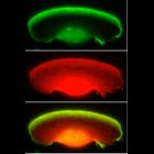
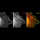
Localization of cross-linking proteins in fibroblast cytoskeleton. Fluorescence ...
CIL:34923
NCBI Organism Classification
Homo sapiens
Biological Process
cellular localization
Cellular Component
lamellipodium
Localization of cross-linking proteins in fibroblast cytoskeleton. Fluorescence ...
CIL:34924
NCBI Organism Classification
Xenopus laevis
Biological Process
cellular localization
Cellular Component
lamellipodium
Localization of cross-linking proteins in fibroblast cytoskeleton. Fluorescence ...
CIL:34925
NCBI Organism Classification
Xenopus laevis
Biological Process
branching of actin filaments
Cellular Component
actin cytoskeleton
Localization of cross-linking proteins in fibroblast cytoskeleton. Immuno-EM of...
CIL:34926
NCBI Organism Classification
Homo sapiens
Biological Process
branching of actin filaments
Cellular Component
actin cytoskeleton
Localization of cross-linking proteins in fibroblast cytoskeleton. Immuno-EM of...
CIL:34927
NCBI Organism Classification
Xenopus laevis
Biological Process
branching of actin filaments
Cellular Component
actin cytoskeleton
Localization of cross-linking proteins in fibroblast cytoskeleton. Immuno-EM of ...
CIL:34928
NCBI Organism Classification
Xenopus laevis
Biological Process
branching of actin filaments
Cellular Component
actin cytoskeleton
Localization of cross-linking proteins in fibroblast cytoskeleton. mmuno-EM of t...
CIL:34929
NCBI Organism Classification
Homo sapiens
Biological Process
branching of actin filaments
Cellular Component
actin cytoskeleton
Localization of cross-linking proteins in fibroblast cytoskeleton. Immuno-EM of...
CIL:34930
NCBI Organism Classification
Xenopus laevis
Biological Process
branching of actin filaments
Cellular Component
actin cytoskeleton
Localization of cross-linking proteins in fibroblast cytoskeleton. Immuno-EM of ...
CIL:35060
NCBI Organism Classification
none specified
Biological Process
branching of actin filaments
Cellular Component
actin cytoskeleton
Electron micrograph of keratocyte or fibroblast lamellipodial actin network afte...
CIL:35061
NCBI Organism Classification
none specified
Biological Process
branching of actin filaments
Cellular Component
actin cytoskeleton
Electron micrograph of keratocyte or fibroblast lamellipodial actin network afte...
CIL:35062
NCBI Organism Classification
none specified
Biological Process
branching of actin filaments
Cellular Component
actin cytoskeleton
Electron micrograph of keratocyte or fibroblast lamellipodial actin network afte...
CIL:35063
NCBI Organism Classification
none specified
Biological Process
branching of actin filaments
Cellular Component
actin cytoskeleton
Electron micrograph of keratocyte or fibroblast lamellipodial actin network afte...
CIL:35064
NCBI Organism Classification
none specified
Biological Process
branching of actin filaments
Cellular Component
actin cytoskeleton
Electron micrograph of keratocyte or fibroblast lamellipodial actin network afte...
CIL:35065
NCBI Organism Classification
none specified
Biological Process
branching of actin filaments
Cellular Component
actin cytoskeleton
Electron micrograph of keratocyte or fibroblast lamellipodial actin network afte...
CIL:35066
NCBI Organism Classification
none specified
Biological Process
branching of actin filaments
Cellular Component
actin cytoskeleton
Electron micrograph of keratocyte or fibroblast lamellipodial actin network afte...
CIL:35067
NCBI Organism Classification
none specified
Biological Process
branching of actin filaments
Cellular Component
actin cytoskeleton
Electron micrograph of keratocyte or fibroblast lamellipodial actin network afte...
CIL:35068
NCBI Organism Classification
none specified
Biological Process
branching of actin filaments
Cellular Component
actin cytoskeleton
Electron micrograph of keratocyte or fibroblast lamellipodial actin network afte...