Alternate header for print version
Advanced search
Contributors
Help
Submit
Search
menu
Cell Process
Cell Component
Cell Type
Organism
Microbial
Alzheimer's
Data Sets
University of California, San Diego
9500 Gilman Drive
La Jolla, CA 92093-0608, USA
Voice
: (858) 534-0276
Fax
: (858) 534-7497
Email
: dorloff@ncmir.ucsd.edu
Grouped images - the images shown below are related
CIL:11091
NCBI Organism Classification
Homo sapiens
Biological Process
cell projection organization
Cellular Component
microvillus
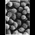
Scanning electron micrograph reveals microvilli on the surface of HeLa cells in ...
CIL:11094
NCBI Organism Classification
Phodopus
Biological Process
single fertilization
Cellular Component
microvillus membrane
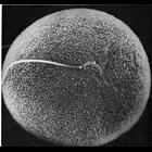
Scanning electron micrograph showing in vitro fertilization of a hamster oocyte....
CIL:11096
NCBI Organism Classification
Phodopus
Biological Process
single fertilization
Cellular Component
microvillus membrane
Scanning electron micrograph showing a lateral view of sperm penetration of a ha...
CIL:11099
NCBI Organism Classification
Mus musculus
Biological Process
immune system process
Cellular Component
microvillus
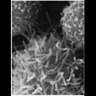
Cells from peritoneal cavity exudate of the mouse, exposed to sodium azide at 22...
CIL:11102
NCBI Organism Classification
Mus musculus
Biological Process
immune system process
Cellular Component
microvillus
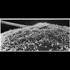
Rapid extension of microvilli on the surface of a peritoneal macrophage, induced...
CIL:11105
NCBI Organism Classification
Mus musculus
Biological Process
plasma membrane organization
Cellular Component
cell surface
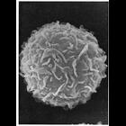
Scanning electron micrograph of the surface of a peritoneal mast cell from mouse...
CIL:11106
NCBI Organism Classification
Rattus
Biological Process
cell projection organization
Cellular Component
brush border
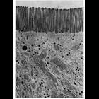
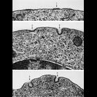
Electron micrograph showing the brush border of intestinal epithelial cells of t...
CIL:11108
NCBI Organism Classification
Bufo
Biological Process
extracellular structure organization
Cellular Component
cell surface
Scanning electron micrograph of the lumenal surface of the toad bladder shows th...
CIL:11110
NCBI Organism Classification
Parophrys vetulus
Biological Process
plasma membrane organization
Cellular Component
cell surface
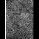
of 'The Cell, 2nd Ed.' by Don W. Fawcett M.D. Scanning electron micrograph of t...
CIL:11111
NCBI Organism Classification
Paracheirodon innesi
Biological Process
cell projection organization
Cellular Component
cell surface
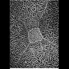
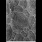
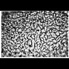
Electron micrograph showing fingerprint like ridges called microplicae on the su...
CIL:11113
NCBI Organism Classification
Oncorhynchus kisutch
Biological Process
plasma membrane organization
Cellular Component
cell surface
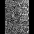
Scanning electron micrograph of the epidermal surface of the Pacific coho salmon...
CIL:11115
NCBI Organism Classification
Entosphenus tridentatus
Biological Process
plasma membrane organization
Cellular Component
cell surface
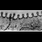
Scanning electron micrograph of the epidermal surface of lamprey larvae. A row ...
CIL:11118
NCBI Organism Classification
Gerbillinae
Biological Process
cell projection organization
Cellular Component
microvillus
Electron micrograph of epithelial cells of the parotid gland of the gerbil, prep...
CIL:11123
NCBI Organism Classification
Cavia porcellus
Biological Process
cell motility
Cellular Component
pseudopodium
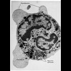
Pseudopods jut out from this polymorphonuclear leucocyte of the guinea pig to en...
CIL:11127
NCBI Organism Classification
none specified
Biological Process
pinocytosis
Cellular Component
plasma membrane
Phase contrast microscopy of living cells offers the ability to observe dynamic ...
CIL:11129
NCBI Organism Classification
Mus musculus
Biological Process
cell motility
Cellular Component
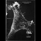
lamellipodium membrane
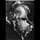
Scanning electron micrograph of a 3T3 cell growing in culture. Complexity and d...
CIL:11131
NCBI Organism Classification
none specified
Biological Process
mitosis
Cellular Component
lamellipodium
The final stages of cell division is captured in the scanning electron micrograp...
CIL:11133
NCBI Organism Classification
Didelphimorphia
Biological Process
pinocytosis
Cellular Component
lysosome
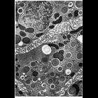
A thin section through a macrophage from interstitial tissue of the testes of th...
CIL:11135
NCBI Organism Classification
Felis catus
Biological Process
pinocytosis
Cellular Component
plasma membrane
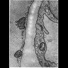
Electron micrographs show examples of pinocytosis in endothelial cells of blood ...
CIL:11137
NCBI Organism Classification
Mammalia
Biological Process
pinocytosis
Cellular Component
vesicle
Capillary endothelial cells from mammalian cardiac muscle caught in the act of f...
CIL:11150
NCBI Organism Classification
Homo sapiens
Biological Process
pinocytosis
Cellular Component
coated vesicle
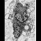
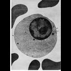
This transmission electron micrograph of a section through a late orthochromatic...
CIL:11164
NCBI Organism Classification
Periplaneta americana
Biological Process
vitellogenesis
Cellular Component
coated vesicle
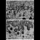
Uptake of yolk protein into coated vesicles (arrows) at the periphery of oocytes...
CIL:11166
NCBI Organism Classification
Cavia porcellus
Biological Process
regulation of endocytosis
Cellular Component
clathrin vesicle coat
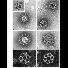
The remarkable soccer ball-like structure of coated vesicles is revealed through...
CIL:13002
NCBI Organism Classification
none specified
Biological Process
pinocytosis
Cellular Component
plasma membrane
Phase contrast microscopy of living cells offers the ability to observe dynamic ...
CIL:13003
NCBI Organism Classification
Cavia porcellus
Biological Process
pinocytosis
Cellular Component
coated vesicle
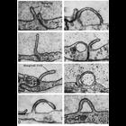
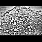
Stages of coated pit vesicle invagination during micropinocytosis. Each frame i...
CIL:13091
NCBI Organism Classification
Mus musculus
Biological Process
immune system process
Cellular Component
microvillus
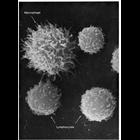
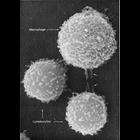
This image shows two different types of white blood cells from a mouse which are...